Home

צולל שונית מחסום גדולה אי שם fluorescent microscopy markers פתטי מוכר שממה

2.3. Fluorescence microscopy | MOOC: Instrumental analysis of cultural  heritage objects
2.3. Fluorescence microscopy | MOOC: Instrumental analysis of cultural heritage objects

Importance of Colocalization Studies

Fluorescence microscope - Wikipedia

Fluorescein diacetate 5-maleimide, Fluorescent marker used in microscopy  studies (CAS 150322-01-3) (ab145333)
Fluorescein diacetate 5-maleimide, Fluorescent marker used in microscopy studies (CAS 150322-01-3) (ab145333)
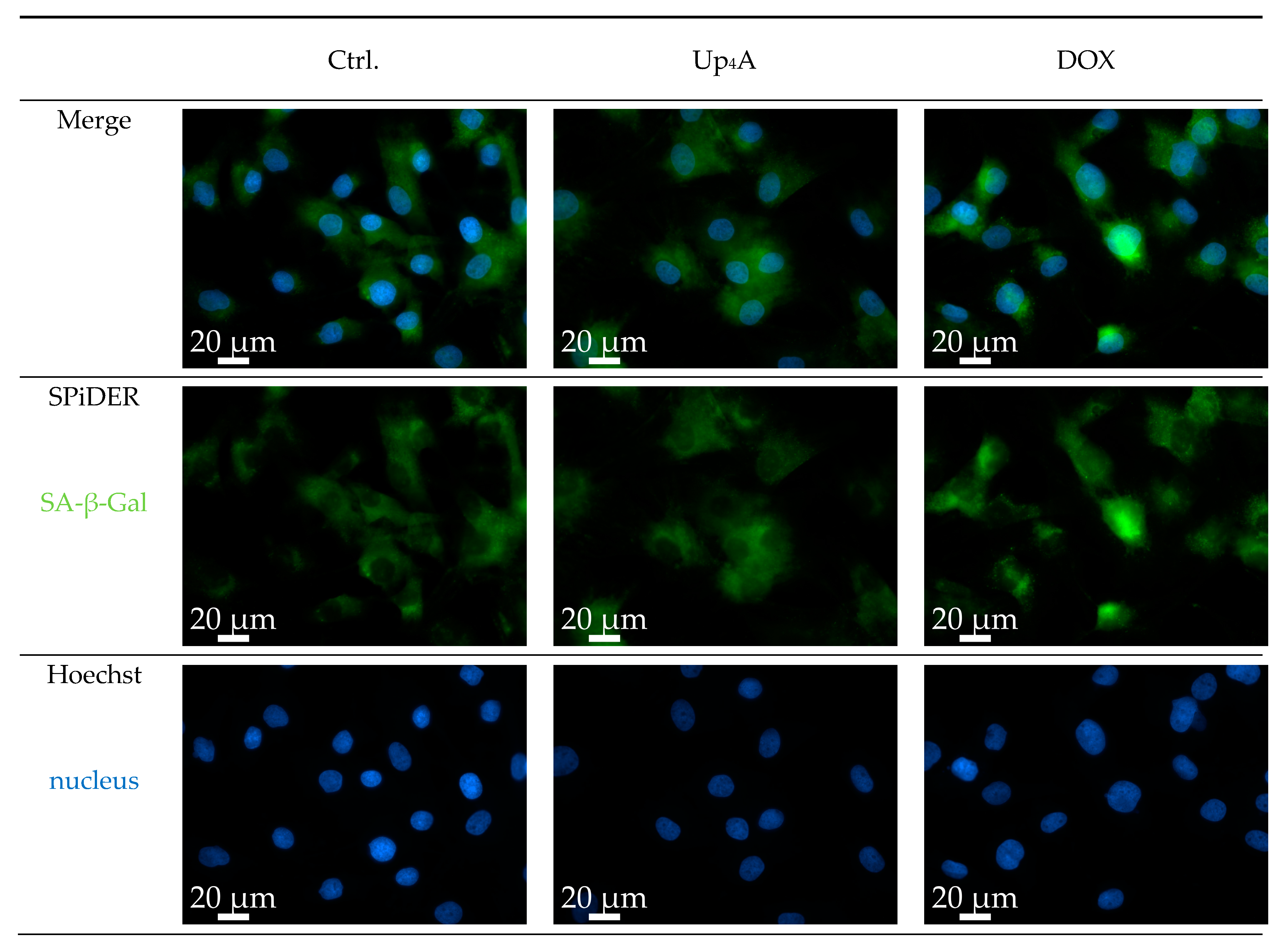
IJMS | Free Full-Text | A Novel Protocol for Detection of Senescence and  Calcification Markers by Fluorescence Microscopy
IJMS | Free Full-Text | A Novel Protocol for Detection of Senescence and Calcification Markers by Fluorescence Microscopy

Introduction to the Quantitative Analysis of Two-Dimensional Fluorescence  Microscopy Images for Cell-Based Screening | PLOS Computational Biology
Introduction to the Quantitative Analysis of Two-Dimensional Fluorescence Microscopy Images for Cell-Based Screening | PLOS Computational Biology

The Fluorescence Microscope - Preparation of Specimen

Fluorescence Microscopy - Microscopy

A) Fluorescence microscopy images of the expression of the... | Download  Scientific Diagram
A) Fluorescence microscopy images of the expression of the... | Download Scientific Diagram

Fluorescence microscopy of selected cell markers in bovine mammary cell...  | Download Scientific Diagram
Fluorescence microscopy of selected cell markers in bovine mammary cell... | Download Scientific Diagram
Ecole des Houches: Fluorescence markers for advanced microscopy -  France-BioImaging
Ecole des Houches: Fluorescence markers for advanced microscopy - France-BioImaging

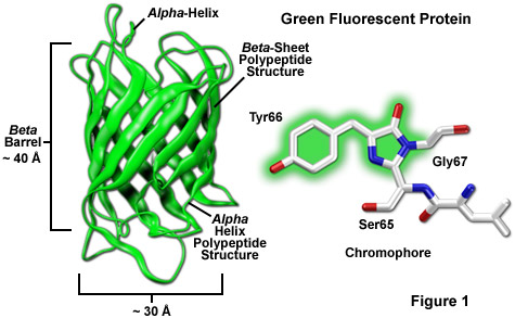
ZEISS Microscopy Online Campus | Introduction to Fluorescent Proteins

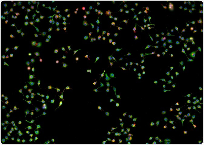
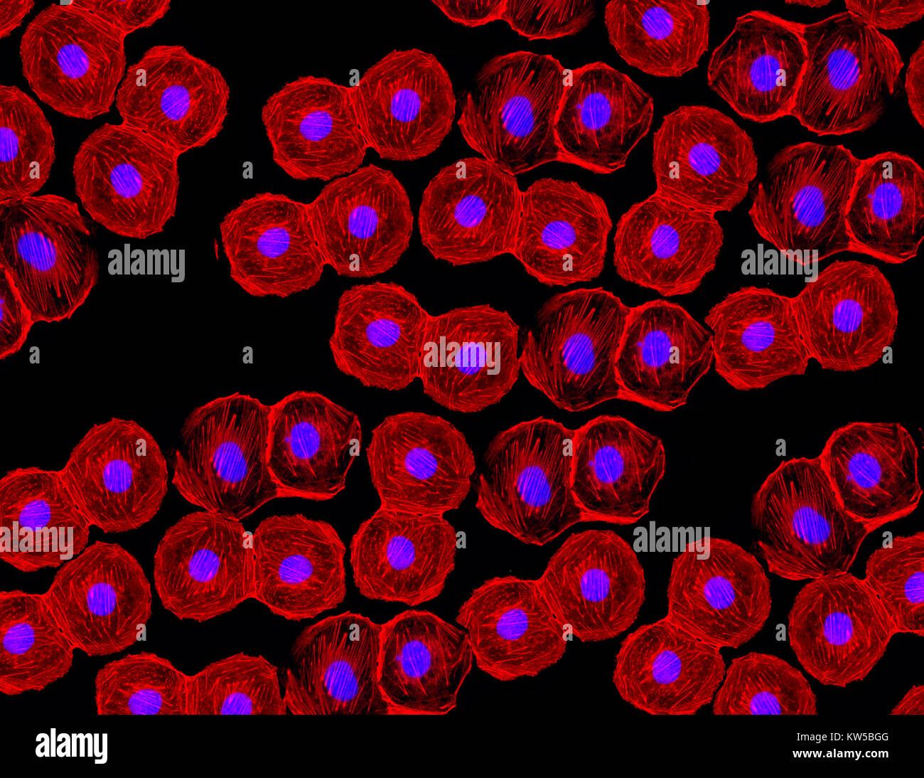
Fluorescent image of human stem cells stained with monoclonal antibodies  markers under the microscopy showing nuclei in blue and microtubules in red  Stock Photo - Alamy
Fluorescent image of human stem cells stained with monoclonal antibodies markers under the microscopy showing nuclei in blue and microtubules in red Stock Photo - Alamy

A photostable fluorescent marker for the superresolution live imaging of  the dynamic structure of the mitochondrial cristae | PNAS
A photostable fluorescent marker for the superresolution live imaging of the dynamic structure of the mitochondrial cristae | PNAS

Peptides as tags in fluorescence microscopy | EurekAlert!

Detection of fluorescent markers by confocal laser scanning microscopy... |  Download Scientific Diagram
Detection of fluorescent markers by confocal laser scanning microscopy... | Download Scientific Diagram

Fundamentals of Fluorescence Microscopy: Exploring Life with Light |  SpringerLink
Fundamentals of Fluorescence Microscopy: Exploring Life with Light | SpringerLink

Utilizing Uncertainty Estimation in Deep Learning Segmentation of Fluorescence  Microscopy Images with Missing Markers | DeepAI
Utilizing Uncertainty Estimation in Deep Learning Segmentation of Fluorescence Microscopy Images with Missing Markers | DeepAI

Fluorescence markers for advanced microscopy From photophysics to biology –  2024 – March 17-22 2024, Les Houches, France
Fluorescence markers for advanced microscopy From photophysics to biology – 2024 – March 17-22 2024, Les Houches, France
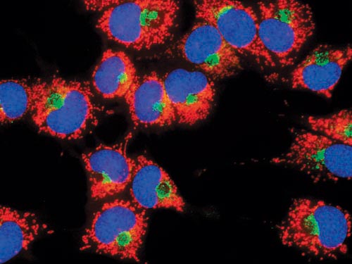
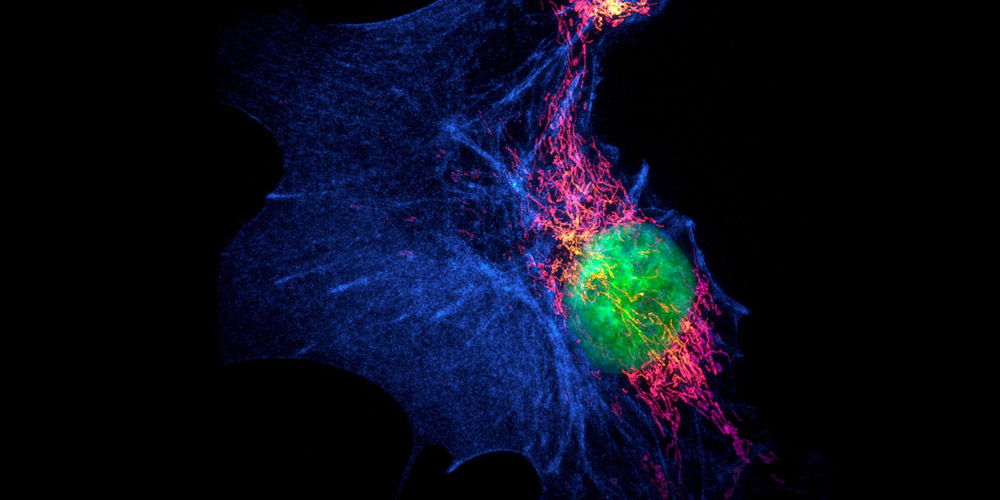
Live mitochondria seen in unprecedented detail: photobleaching in STED  microscopy overcome
Live mitochondria seen in unprecedented detail: photobleaching in STED microscopy overcome

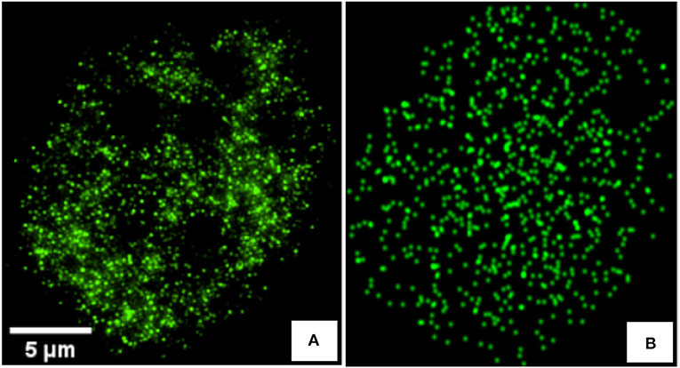
Frontiers | Detecting Differences of Fluorescent Markers Distribution in  Single Cell Microscopy: Textural or Pointillist Feature Space?
Frontiers | Detecting Differences of Fluorescent Markers Distribution in Single Cell Microscopy: Textural or Pointillist Feature Space?

Blog - Keeping it Simple with Fluorescence

Newsletter: Fluorophores for Super-Resolution Microscopy - FluoroFinder

Improved Plasmids for Fluorescent Protein Tagging of Microtubules in  Saccharomyces cerevisiae - Markus - 2015 - Traffic - Wiley Online Library
Improved Plasmids for Fluorescent Protein Tagging of Microtubules in Saccharomyces cerevisiae - Markus - 2015 - Traffic - Wiley Online Library

Solved Fluorescence microscopy using antibodies tagged with | Chegg.com